伊人直播
The traditional view holds that nano-droplets are unstable due to their high surface energy. However, Professor HuanWangʼs research group observed the formation process of nano-droplets using liquid-phase electron microscopy (LP-EM), influenced by confinement and surface forces. In a graphene-encapsulated liquid cell, nanobubbles acted as geometric constraints, leading to the formation of a thin water film approximately 10nm thick. They found that nano-droplets can indeed form and growstably. The droplet formation is primarily driven by Plateau-Rayleigh instability, and the droplets' stability is influenced by geometric confinement. The experiments' newly discovered "liquid bridge" mechanism mainly helps stabilize the nano-droplets. These observations provide new insights into nanofluidics and arepotentially significant for understanding and utilizing fluid behavior in confined environments. Moreover, they also offer a research method to explore new phenomena in chemical reactions within micro- and even nano-droplets. (J. Phys. Chem. B 2024, 128, 3732)
To explore nanoscale self-assembly and non-equilibrium droplet evaporation dynamics, HuanWang's research group, in collaboration with Yuanhua Shao's research group, developed a quartz nanopipette liquid cell. This new liquid cell, with adjustable size and a nano opening, allows observation of evaporation phenomena in a vacuum. It can also be sealed to become a closed liquid cell, serving as a low-cost alternative to mainstream silicon nitride cells. The coffee ring effect, a common physical phenomenon during the evaporation of micron-sized droplets, involves capillary flow driven by surface tension and evaporation rate gradients. This flow transports colloidal particles to the edges, forming rich and ordered patterns. This phenomenon has a potential impact on materials science, chemistry, and physics. Due to the lack of direct imaging methods, confirming its existence at the nanoscale remains challenging. The key question is whether the flow field can overcome the strong thermal fluctuations at the nanoscale for directed assembly and pattern formation. This study tracked nanoparticles within an evaporating nano-droplet, observing the nano coffee ring phenomenon and quantifying the particle motions during the process (Fig. 1).
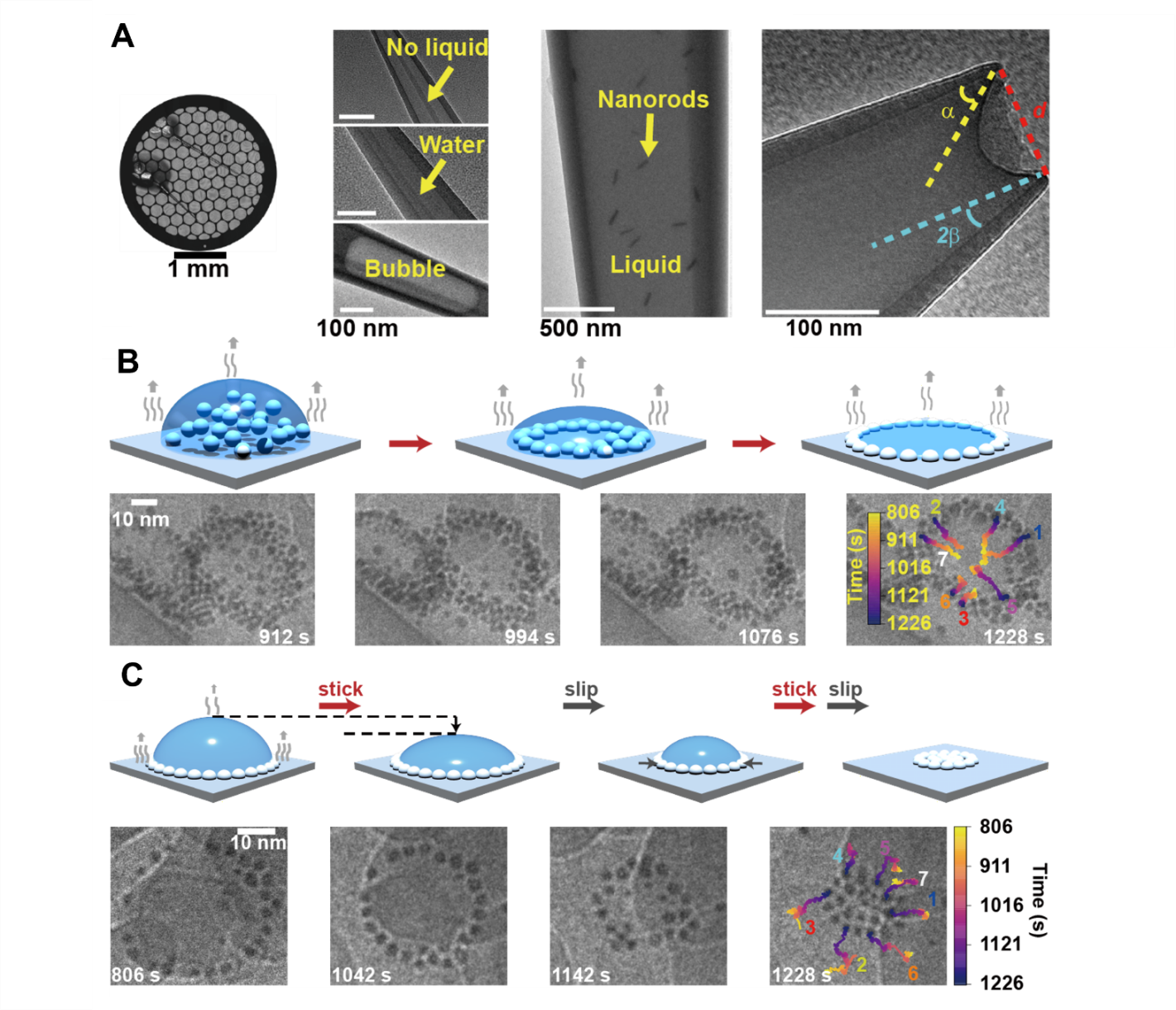
Fig.1 A: Optical microscope image of the nanopipettes placed on the grid; electron microscope images of the nanopipettes showing the absence of liquid, fully filled with liquid, presence of bubble, presence of nanorod particles, and the concave liquid surface at the tip. B: Formation of the nano coffee ring. C: Reversal of the nano coffee ring.
This work reports a quartz nanopipette liquid cell electron microscopy imaging technique that allows direct imaging of both open and closed systems. This advancement democratizes the use of LP-EM imaging,making it a practical method for general users without the need for specialized operators or high costs. Direct imaging revealed the significant influence of capillary flow within tens of nanometers in evaporating nano-droplets. During the formation of nano coffee rings, the rich contact line dynamics highlighted the importance of the interactions between nano-flow, surface forces, and thermal fluctuations in guiding pattern formation.
This work, titled "Nanopipette dynamic microscopy unveils nano coffee ring", was published in the Proceedings of the National Academy of Sciences of the United States of America(direct submission). The corresponding authors are Huan Wang and Yuanhua Shao from the College of Chemistry and Molecular Engineering at Peking University. The first authors are Ph.D. student Deyi Zhang (Huan Wang group) and Dr. Yi Shao (Yuanhua Shao group). Professor Sheng Mao from the College of Engineering at Peking University, Professor Jingjing Wei from the School of Chemistry and Chemical Engineering at Shandong University, Professor Limin Qi (Ph.D student: Jiayi Zhou) from the College of Chemistry and Molecular Engineering at Peking University, and related team members have made significant contributions to this work. This research was supported by the National Natural Science Foundation of China, the National Biomedical Imaging Center at Peking University, and the Spectroscopy Center at Peking University, and received assistance from the Electron Microscopy Laboratory and the Analytical Instrumentation Center at Peking University.
Link to the original article: //www.pnas.org/doi/10.1073/pnas.2314320121